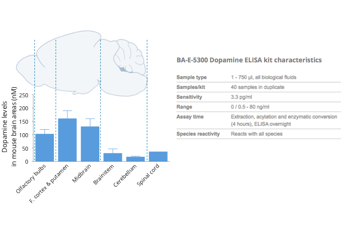
Figure 1: Mouse brain tissue levels of dopamine quantified and shown in different areas using a specific dopamine ELISA kit (BA-E-5300). As expected, high levels of dopamine were detected in the afferent and efferent dopaminergic systems, i.e. putamen, midbrain, and olfactory bulbs.
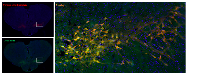
Figure 2: Dopaminergic nigral neurons in coronal rat brain sections revealed by anti-dopamine (DA, rabbit polyclonal antibody #IS1005) and Tyrosine Hydroxylase (TH, #MAB5280, reference catecholaminergic neuron immunostaining). As highlighted in the overlay, DA immunoreactivity localizes with TH staining in the Substantia nigra pars compacta, thus showing that our antibody against the DA neurotransmitter – used with the STAINperfect immunostaining kit A – is a validated tool to directly highlight DAergic system networks.


Offer valid until February 25, 2020