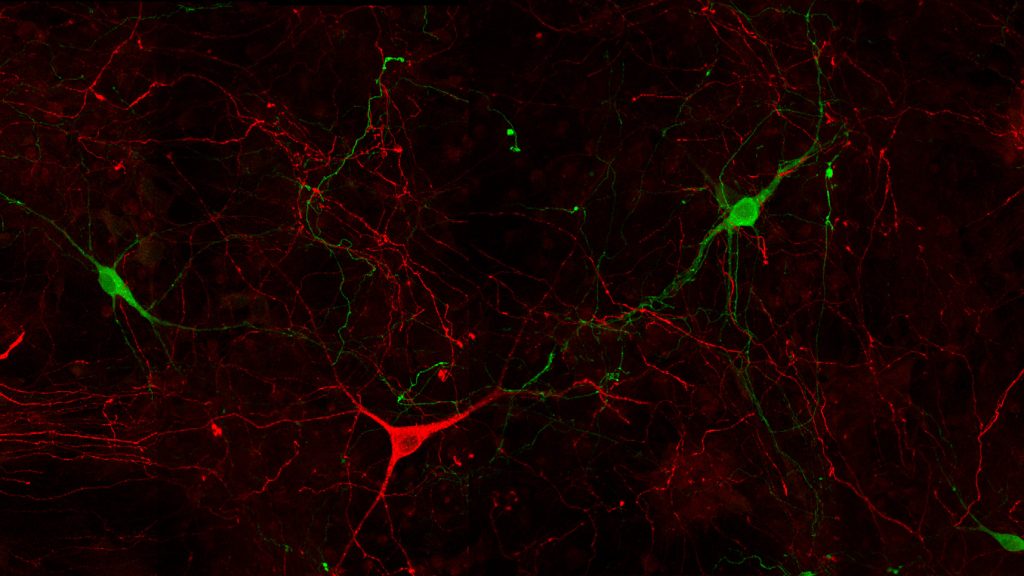
[vc_row][vc_column][vc_single_image image=”14065″ img_size=”full” onclick=”custom_link” link=”https://preprod.immusmol.com/shop/serotonin-goat-pab/”][vc_column_text]Dopamine (green) & Serotonin (red) in mouse primary neuronal culture using STAINperfect immunostaining kit with IS1005 (DA pAb) and IS1035 (5-HT pAb)[/vc_column_text][vc_empty_space][vc_column_text]Dopamine, Serotonin, GABA, Glutamate, Histamine, Norepinephrine, Serine, Octopamine, … To generate spatial information about key neuromodulators and neurotransmitters, neurobiologists typically perform stainings against enzymes (e.g. TH for dopamine, GAD65-67 for GABA), receptors (e.g. Glutamate) or transporters (e.g. Norepinephrine, Glutamate). Why? Simply because highly specific antibodies against neuromediators weren’t available until recently. [/vc_column_text][vc_column_text]Our team developed and IF/IHC-validated 17 primary antibodies to key neurotransmitters. These antibodies were already used in 20+ papers. Take a look below. And for easy quantification, see our ELISA kits to neurotransmitters.[/vc_column_text][vc_column_text]
[/vc_column_text][vc_row_inner][vc_column_inner][vc_column_text]
[/vc_column_text][vc_single_image image=”14995″ img_size=”full” alignment=”center” onclick=”custom_link” link=”https://preprod.immusmol.com/shop/dopamine-rabbit-pab/”][vc_column_text]Immunostaining in the CNS of embryonic mouse E13.5 using STAINperfect kit and primary antibodies to Dopamine (IS1005 – green) and Serotonin (IS1035 -red)[/vc_column_text][/vc_column_inner][/vc_row_inner][vc_row_inner][vc_column_inner][vc_column_text]
[/vc_column_text][vc_single_image image=”15005″ img_size=”full” alignment=”center” onclick=”custom_link” link=”https://preprod.immusmol.com/shop/gaba-rabbit-pab/”][vc_column_text]Immunodetection of MAP2 (red) and GABA (green) mouse primary cortical cultures, using anti-GABA IS1006 antibody and the STAINperfect immunostaining kit.[/vc_column_text][/vc_column_inner][/vc_row_inner][vc_row_inner][vc_column_inner][vc_column_text]
[/vc_column_text][vc_single_image image=”15007″ img_size=”full” alignment=”center” onclick=”custom_link” link=”https://preprod.immusmol.com/shop/l-glutamate-rabbit-pab/”][vc_column_text]Immunodetection of L-Glutamate (green) and MAP2- (red) positive neurons in mouse primary cortical culture reveals the presence of L-Glutamate within fibers and soma of neurons (IS1001 anti-glutamate rabbit pAb)[/vc_column_text][/vc_column_inner][/vc_row_inner][vc_row_inner][vc_column_inner][vc_column_text]
[/vc_column_text][vc_single_image image=”15008″ img_size=”full” alignment=”center” onclick=”custom_link” link=”https://preprod.immusmol.com/shop/l-serine-pab/”][vc_column_text]Primary mouse cortical neurons were stained using anti-L-Serine (green) polyclonal rabbit antibody (IS1003) and anti-GABA (red) chicken polyclonal antibody (IS1036) using the STAINperfect immunostaining kit A.[/vc_column_text][/vc_column_inner][/vc_row_inner][vc_row_inner][vc_column_inner][vc_column_text]
[/vc_column_text][vc_single_image image=”15009″ img_size=”full” alignment=”center” onclick=”custom_link” link=”https://preprod.immusmol.com/shop/octopamine-rabbit-pab/”][vc_column_text]Immunostaining of crayfish eyestalk using anti-octopamine rabbit polyclonal antibody (green) and anti-serotonin goat polyclonal antibody (red). Tissues were processed with whole mount protocol of STAINperfect kit A.[/vc_column_text][/vc_column_inner][/vc_row_inner][vc_row_inner][vc_column_inner][vc_column_text]
[/vc_column_text][vc_single_image image=”15010″ img_size=”full” alignment=”center” onclick=”custom_link” link=”https://preprod.immusmol.com/shop/serotonin-polyclonal-antibody-is1086/”][vc_column_text]Rabbit polyclonal antibody IS1086 highlights the presence of 5-HT enterochromaffin cells of the intestinal mucosa.[/vc_column_text][/vc_column_inner][/vc_row_inner][vc_column_text]
Our team developed 17 antibodies to key neurotransmitters, together with a dedicated immunostaining kit for straightforward multiplexed IF/IHC imaging in cell cultures, whole mounts, and tissue sections.
A trial pack is available, containing the STAINperfect staining kit and 2 antibodies.
If you have any questions about staining protocols or sample preparation, ask us ! Our antibodies were used in 20+ papers to stain stem cell-derived midbrain organoids, spider tissues, human brain tissues, and more. So we might have answers for you!
And to quantify neurotransmitters in supernatant and biological samples, we developed highly sensitive ELISA kits, cross-validated by LC-MS, which have already been cited in 100+ papers.
[/vc_column_text][/vc_column][/vc_row]
